SBAA569 November 2022 AFE4960 , AFE4960P
1 Application Brief
Application
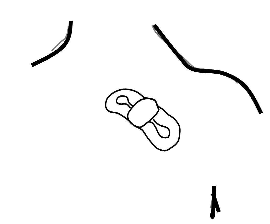
Remote Patient Monitoring (RPM) refers to the use of remotely-accessible sensors to monitor the vital signs of patients both within and outside conventional clinical settings, for example, from the home. An example is the use of a skin patch (see Figure 1-1) for the continuous and long-term (usually several days) monitoring of electrocardiograms (ECG) to detect heart arrhythmias like atrial fibrillation. The functionality required in such monitoring devices can include any combination of the following; single- or multi-lead ECG, respiration using thoracic impedance measurement, temperature, blood pressure, or SpO2 monitoring. The AFE4960 device brings an unmatched level of integration for remote patient monitoring applications, and enables a single-chip realization of a 3-lead ECG system. The device supports 2-channel ECG, respiration, and automatic pacemaker detection. Figure 1-2 shows the connection of the AFE4960 pins to electrodes of a 3-lead ECG. The electrodes marked as LA, RA, LL, and RL refer to the Left and Right limb (Arm, Leg) electrodes. The electrodes take their notations from clinical ECG systems though their positions can be different in an RPM device. Figure 1-3 shows a reference schematic for a 3-lead ECG system using AFE4960. The AFE4960P additionally contains a photoplethysmography (PPG) signal chain that enables high-accuracy SpO2 measurement. Figure 1-4 shows a reference schematic for a 3-lead ECG + SpO2 system using the AFE4960P. The AFE4960 and AFE4960P are small devices with low power consumption. These features make these devices excellent choices for a variety of RPM devices such as wearable Holter monitors and bio-patches.
AFE4960, AFE4960P- AFE4960: 2-channel ECG, respiration, pace detect
- AFE4960P: AFE4960 + PPG signal chain
- Package: 2.6-mm × 2.6-mm DSBGA, 0.4-mm pitch
- Supply: RX: 1.7 V–1.9 V; TX: 3 V–5.5 V (for PPG)
- Interface: SPI™, I2C interfaces, first in, first out (FIFO) with 128-sample depth
 Figure 1-1 Bio-patch, an Example of an RPM
Device
Figure 1-1 Bio-patch, an Example of an RPM
DeviceDifferentiation
- 2-channel ECG signal acquisition from small form-factor electrodes with high contact impedance – high input impedance, right leg drive (RLD) electrode to improve common-mode rejection ratio (CMRR)
- Integrated low-pass filter (LPF) filters high-frequency noise
- AC, DC lead detect and lead impedance measurement
- Low-noise respiration signal chain
- Automatic pace detection at 20-μA extra current
- AFE4960P: PPG acquisition enables SpO2. Synchronized acquisition of PPG and ECG signals enables pulse transit time (PTT) based blood pressure estimation
Table 1-1 lists the key specifications for the 3-lead ECG + SpO2 system.
| Parameter | AFE4960P | Comments |
|---|---|---|
| Number of electrodes | ECG channels | 4 electrodes | 2 ECG channels | Enables a single-chip design for a 3-lead ECG |
| ECG input referred noise | 5 μVPP | In 150-Hz bandwidth |
| ECG channel CMRR | 130 dB | With RLD electrode driven through feedback loop |
| Current consumption | 222 μA per channel | Per ECG channel at 500-Hz sampling rate |
| Respiration impedance accuracy | 40 mΩPP | Over a bandwidth of 0.05 Hz to 2 Hz while measuring 2-kΩ baseline |
| Pace detection – minimum amplitude | width | 2 mV | 100 μs | None |
| SNR of PPG signal chain | 110 dB | High SNR enables high-accuracy SpO2 monitoring even for cases of low perfusion index |
Figure 1-5 shows the AFE4960 signal chain interfacing with the four electrodes of a 3-lead ECG system. Channel 2 can be configured as either a second ECG channel or a respiration impedance channel. An automatic pace detection can also be enabled on the lead connected to channel 2.