SSZTAA7 march 2017 AFE2256
Diagnostic imaging technologies like X-ray systems are continuously evolving to improve not just the quality of patient diagnostics but also clinical efficiency. The ability to accurately and cost-effectively capture detailed images of the human body to achieve these goals is becoming increasingly critical across small to large modern medical practices, due to the market becoming more competitive. Additionally as the market becomes more competitive, there erupts numerous advancements in the technology.
Introduced in the mid-1990s, X-ray systems capture higher-quality images at higher resolutions than ever before, all while requiring less time to capture. Digital X-ray systems have revolutionized diagnostic radiology. Many of the improvements in X-ray systems are a direct result of the advantages of digital capture over traditional film capture.
Digital X-ray systems have a higher dynamic range than film, which allows for clearer, more detailed images. Since digital X-ray systems do not require extensive processing, there is a significant reduction in the time it takes to deliver images to radiologists and ultimately patients. Digital capture also improves image-processing capabilities such as spatial zooming and contrast enhancement. Finally, there are more convenient storage options for digital images than film, and digital storage reduces the amount of polluting waste products.
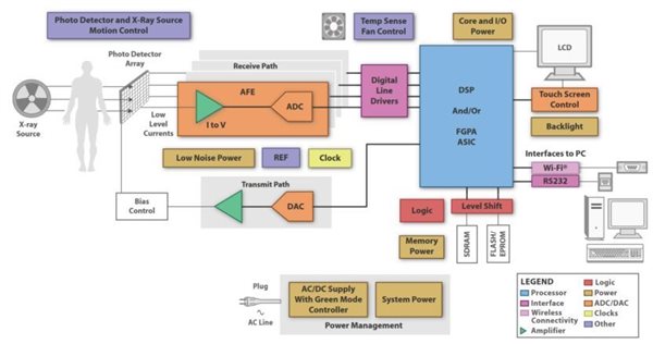
Given all of the improvements over the years, X-ray systems have become very complex and require an analog front end (AFE), a digital signal processor (DSP), an LCD, power supply design, sensor controls and a few other external components. The block diagram in Figure 1 shows the readout electronics required for direct imaging to convert a charge in a flat panel detector (FPD) to digital data. There are two chains: the acquisition chain and the biasing chain. At the start of the acquisition chain, an AFE circuit (sometimes called the readout integrated circuit) digitizes the charges from the array of pixels on the FPD. The biasing chain generates bias voltages for the thin film transistor (TFT) array through intermediate bias and gate control circuitry. A DSP, field-programmable gate array (FPGA), application-specific integrated circuit (ASIC) or a combination of these applies signal conditioning. These processors also manage high-speed serial communications with the external image-processing unit through a high-speed interface.
 Figure 1 X-ray System
Devices like the AFE2256 AFE are specifically designed for FPD-based digital X-ray systems. This device can be used for static or dynamic imaging and also features multiple sleep and power-down modes that are especially useful for battery-powered systems. The AFE2256 has 256 channels and an onboard 16-bit ADC.
With the evolution of X-ray systems, there is an increasing demand for clearer, more detailed images, faster scan times, better storage options and simpler power-supply schemes. The ability to accurately and cost-effectively achieve these goals becomes easier with a device like the AFE2256. There are a few key differentiators of this device to others on the market. The AFE2256 is very easy to use; there is a simple power supply management scheme enabling smaller board space and less external circuitry. This device also has low power consumption, especially at low integration times, which enables longer battery life. There is also low noise: 750 electrons RMS with 1.2pC input charge range, which enables lower dosage and dynamic imaging resulting in clearer images. Faster scan times (less than 20s) also allow for more efficient image captures.
TI has a long history and continually invests in digital X-ray technology. To learn more, check out our entire X-ray portfolio, including the AFE2256.
Additional Resources
- Download the AFE2256 data sheet.