TIDUDO6B May 2019 – October 2020
- Description
- Resources
- Features
- Applications
- 5
- 1System Description
-
2System Overview
- 2.1 Block Diagram
- 2.2 Highlighted Products
- 2.3
System Design Theory and Design Considerations
- 2.3.1 AFE4900 and Power Supply
- 2.3.2 CC2640R2F Microcontroller
- 2.3.3 PPG Measurement
- 2.3.4 ECG Measurement
- 2.3.5 Selecting TX Supply (TX_SUP) Value for Driving LEDs
- 2.3.6 Generating TX Supply for Driving LEDs
- 2.3.7 Generating RX Supply for AFE4900
- 2.3.8 Generating I/O Supply
- 2.3.9 Battery Input and Reservoir Capacitors
- 2.3.10 Battery Life Calculations
- 2.3.11 External Memory
- 2.3.12 LED Indications
- 2.3.13 Connections Between Sensor Board and ECG Board
-
3Hardware, Software, Testing Requirements, and Test Results
- 3.1 Required Hardware and Software
- 3.2 Testing and Results
-
4Design Files
- 4.1 Schematics
- 4.2 Bill of Materials
- 4.3
PCB Layout Recommendations
- 4.3.1 Layout for Main Board
- 4.3.2 Connection From PDs to AFE
- 4.3.3 Connections From LEDs to AFE
- 4.3.4 Connections From ECG PADs to AFE
- 4.3.5 Connections Between BT and AFE
- 4.3.6 Connections Between BT Antenna and Chip
- 4.3.7 Boost Converter
- 4.3.8 Buck-Boost Converter
- 4.3.9 Layouts for PPG Sensor Boards
- 4.3.10 Layout for ECG Sensor Board
- 4.3.11 Layout Prints
- 4.4 Altium Project
- 4.5 Gerber Files
- 4.6 Assembly Drawings
- 5Software Files
- 6Related Documentation
- 7About the Authors
- Revision History
1.1 Introduction to Parameters Measured Using TIDA-01580
The TIDA-01580 device can be used to measure the following parameters: ECG, PPG, HRM, SpO2, and PTT.
ECG is an electrical measurement of the activity in the heart whereas PPG is an optical measurement of the volume of an organ. In principle, ECG uses multiple electrodes to measure the electrical activity of the heart, whereas PPG illuminates the skin and subcutaneous tissue with light of a specific wavelength from a light-emitting diode (LED) to measure organ volume. This light is absorbed, passed through, or reflected back. A photodiode sensor measures the light that is either transmitted or reflected, depending on where it is placed relative to the LED. The light is then converted to an electrical signal. In both cases, the information can be used to determine the heart rate of a person, but each application offers its own set of diagnostic information. ECG focuses on the electrical activity of the cardiac muscle tissue, because the exact sequence of contraction is well-known to trained cardiologists. Physicians use ECG to diagnose all kinds of heart diseases and abnormalities.
PPG provides more information about blood flow and blood pressure. This measurement can be conducted at various locations on the body, to examine the blood flow to different regions. When measured closest to the aorta of the heart (for example, the left arm), some additional information can be gained regarding the cardiac output and heart valve function. One advantage that PPG has is the number of skin contacts which are required to measure it. Because users can determine PPG from reflected or transmitted light, only a single point of contact is necessary to measure it. This feature allows for easy, continuous, time measurements, which is the most attractive advantage to wearable electronics such as fitness trackers. In contrast, ECG requires that the potential be measured across the heart. This means that users need at least two points of contact: positive and negative. Typically, this connection is only made for a finite period of time.
Figure 1-1 shows the typical waveforms for ECG (blue) and PPG (red). The time difference between the R-peak in the ECG waveform and the arrival of the blood pressure wave in the extremities is another measurement known as PTT. PTT involves simultaneous measurement of ECG and PPG.
 Figure 1-1 Introduction to ECG, PPG, and PTT
Because the frequency of both ECG and PPG signals is same, heart rate can be calculated using both waveforms. Mostly green LEDs are used for measuring heart rates. A green LED has often been used in the reflective sensor to extract the PPG signal. Due to its wavelength, green light is known to penetrate the tissue less than higher wavelength LEDs. Hence, more unabsorbed (reflected) light comes out of the tissue with green than with other colors. Sensing the green light from more than one PD or eliminating from more than one LED surrounding the PD helps.
Red and infrared (IR) lights are used for pulse oximetry, to estimate the true hemoglobin oxygen saturation of arterial blood. Oxyhemoglobin (HbO2) absorbs visible and infrared IR light differently than deoxyhemoglobin (Hb), and appears bright red as opposed to the darker brown of Hb. Absorption in the arterial blood is represented by an AC signal that is superimposed on a DC signal, representing absorptions in other substances like pigmentation in tissue, venous, capillary, bone, and so forth. The cardiac-synchronized AC signal is approximately 1% of the DC level. This value is referred to as the perfusion index %. Equation 1 approximates the ratio of ratios, R, and % SpO2 is calculated as follows:
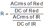
Equation 2 gives the standard model of computing SpO2. This model is often used in this literature in the context of medical devices. However, accurate % SpO2 is computed based on the empirical calibration of the ratio of ratios for the specific device.